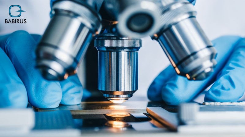
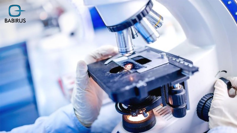
Techniques Used in Cytopathology Microscopy

In cytopathology microscopy, each cell reveals a story waiting to be uncovered, thus, using specialized microscopy techniques will help cytopathologists analyze even the smallest cellular structures to catch diseases early, sometimes before they fully develop.
Moreover, cytopathology microscopy can transform the invisible into the visible with varied techniques that allow pathologists to examine cellular structures, identify abnormalities, and make critical diagnostic decisions.
Read on to explore the essential and emerging methods and techniques in cytopathology microscopy, including light microscopy, immunohistochemistry, and digital image analysis, to learn how these tools are effectively advancing cellular diagnostics.
The Importance of Microscopy in Cytopathology:
Microscopy is a cornerstone of cytopathology, simply because applying different cytopathology microscopy techniques allows specialists to analyze cell structure, protein expression, and other cellular details to detect cancers, infections, and various conditions at early stages.
In other words, cytopathology microscopy allows pathologists to study cells at high magnification and spot delicate abnormalities that might signal disease.
With all the medical technology advancement, the microscopy methods continue to develop as well with broadened accuracy levels and new cytological diagnostics techniques:
· Light Microscopy:
Light microscopy is one of the most advanced tools in cytopathology, with quality results that provide pathologists with an opportunity to observe the cell structure and morphology.
o Brightfield Microscopy:
We can say that brightfield microscopy is the traditional and most widely used form of light microscopy.
This method is ideal for routine cytological analyses, it works by passing white light directly through a stained specimen to improve contrast and reveal cellular details, making it perfect for identifying cancer cells or inflammation.
o Phase-Contrast Microscopy:
This technique is amazing for observing live cells and tracking cellular processes in real time as it boosts the visibility of transparent, unstained samples by strengthening small differences in light as it passes through various parts of the cell.
Moreover, this technique is valuable due to its ability to preserve delicate or unstained samples that might be damaged by dyes, and it can provide early clues to abnormal cell behavior by revealing subtle features like organelles.
o Polarized Light Microscopy:
Polarized light microscopy is highly useful for spotting crystals or amyloid deposits, aiding in the diagnosis of conditions like gout or metabolic disorders where crystals form within tissues.
Especially that this technique uses polarized light to highlight birefringent (light-refracting) structures within cells, making it easier to identify crystalline or fibrous materials.
Immunohistochemistry (IHC):
When talking about cytopathology microscopy, then we have to talk about immunohistochemistry (IHC), which is an invaluable tool for cellular diagnostics that works based on specialized microscopy techniques to identify specific proteins within cells based on specific used antibodies.
Furthermore, the IHC enables cytopathologists to visualize markers that indicate disease by binding antibodies to targeted proteins, moreover, it is widely used in cancer cases due to its ability to detect tumor markers, like HER2 in breast cancer or PSA in prostate cancer, helping to differentiate between tumor types and evaluate their aggressiveness.
So, we can say that IHC provides a high level of precision, which allows pathologists to classify tumors more accurately and even guide treatment options based on the observed protein expression.
The application of IHC provides quicker and more reliable infection detection, aiding clinicians in making timely and informed treatment decisions, which is highly important in diagnosing infectious diseases by marking specific viral or bacterial antigens within tissue samples.
Fluorescence Microscopy:
Fluorescence microscopy is an advanced technique in cytopathology that uses fluorescent dyes to label specific cellular components, allowing cytopathologists to examine multiple structures within a single cell.
These dyes release light under certain wavelengths, making proteins, nucleic acids, and other molecules visible with their remarkable details.
All of that makes this technique in cytopathology vital for identifying distinct cell types, studying protein interactions, and spotting abnormal markers often linked to cancer and other diseases.
Thus, the use of fluorescence microscopy is important for identifying cancer and detecting markers related to autoimmune diseases, infections, and genetic disorders, giving cytopathologists a useful tool for analyzing unique cellular features.
On the other hand, from a research perspective, fluorescence microscopy aids in expanding our knowledge of cellular processes in health and disease, studying cell behavior, and supporting drug development.
Confocal Microscopy:
Confocal microscopy delivers high-resolution and three-dimensional images of cellular structures, moreover, this advanced technique is developed based on fluorescence microscopy.
Additionally, this cytopathology microscopy technique uses laser scanning to capture and analyze cells layer by layer, making it especially valuable in cytopathology for examining tissue samples with complex cell arrangements.
In cytopathology, confocal microscopy is ideal for analyzing thick or densely packed tissue samples, allowing cytopathologists to examine intricate cellular patterns without distortion, especially since it provides sharp, clear images that improve diagnostic accuracy.
Furthermore, all these provided details of cellular architecture help pathologists detect even the most delicate differences between healthy and diseased tissues.
One more thing, confocal microscopy plays a vital role in research, as it supports studies of cell morphology, interactions, and spatial orientation, allowing for improved treatments in clinical cytopathology.
Digital Image Analysis:
Digital image analysis is a modern breakthrough in cytopathology microscopy, particularly in that it combines microscopy with digital technology to improve the accuracy and efficiency of cellular diagnosis.
With this method, cytopathologists can capture, store, and access high-resolution digital images, based on advanced software algorithms, of cell samples for both immediate analysis and future reference, allowing accurate measurement and quantification of cellular details, including size, shape, and color intensity.
This digital approach not only boosts the diagnostic process and provides quicker and more standardized results, but also helps identify subtle abnormalities, detect specific markers, and assess sample quality, boosting diagnostic accuracy.
Moreover, digitalization relies on advanced systems, like LTS-BL1000 from Lituo Biotechnology, which allow and facilitate the managing of large sample volumes, enabling remote image sharing, specialist consultations, and access to comprehensive databases for comparison.
Emerging Microscopic Techniques:
The great innovative advances in cytopathology microscopy are opening the way with new techniques offering unprecedented insight into cellular details for analysis and studie:
- Super-resolution microscopy expands the resolution limits of traditional light microscopy, allowing cytopathologists to observe molecular structures, which is highly valuable in cancer and genetic research, where seeing cellular markers at such depth is crucial.
- Multiplex fluorescence microscopy, another emerging method, enables the visualization of multiple targets within a single cell, helping pathologists analyze complex interactions, particularly within tumors.
- Raman spectroscopy, which captures a cell’s unique molecular “fingerprint” and detects disease-related biochemical changes without staining, and cryogenic electron microscopy (Cryo-EM), which preserves cells in their natural state for detailed views of structures like viruses and proteins.
- Atomic Force Microscopy (AFM), which examines the cell surface topography, and Digital Holographic Microscopy (DHM), which creates 3D images of live cells, support more precise cellular analysis.
Together, these emerging techniques in cytopathology are expanding the potential for early disease detection and enabling more customized diagnostic approaches.
FAQs About Cytopathology Microscopy:
It is time to share with you the most asked question about cytopathology microscopy with reliable answers from our professionals:
· What Types of Microscopes Are Used in Cytopathology?
Cytopathologists use a wide range of microscopes based on the specific analysis needed, including light, fluorescence, and confocal microscopes.
· How Does Immunohistochemistry Help in Cytopathology?
Immunohistochemistry is used to identify particular proteins within cells, making it essential for precise cancer diagnosis by detecting tumor markers and assessing protein expression.
· What Is the Difference Between Brightfield and Phase-Contrast Microscopy?
Brightfield microscopy uses standard white light for viewing stained samples.
While phase-contrast microscopy improves contrast in unstained samples, making it ideal for examining live cells.
· What Are the Advantages of Confocal Microscopy in Cytopathology?
Confocal microscopy offers great features, including high-resolution and three-dimensional images that allow for a detailed look at cellular structures, improving the accuracy of cytopathology analyses.
· How Can Digital Image Analysis Improve the Accuracy of Cytological Diagnosis?
Digital image analysis provides pathologists with information and insight to measure cellular features precisely and capture high-resolution images for detailed review, which helps make diagnoses faster and more reliable.
· What Are the Emerging Trends in Super-Resolution Microscopy for Cytopathology?
Super-resolution microscopy is evolving cytology by permitting visualization of cellular structures at the molecular level, providing more details and insights than traditional microscopy methods.
· How Can Microscopy Techniques Be Combined with Other Diagnostic Methods?
Microscopy can be integrated with molecular diagnostics, like PCR, or paired with imaging techniques to offer a more comprehensive view of cellular abnormalities.
To conclude,
Cytopathology microscopy is at the core of cellular diagnosis, with advanced used tools and techniques to visualize cells with clarity and precision.
Moreover, there are always new technologies emerging in the field of cytopathology to help cytopathologists provide accurate diagnoses with early and effective disease detection.

