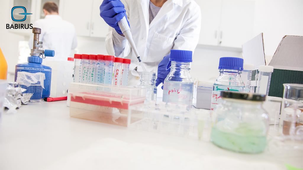
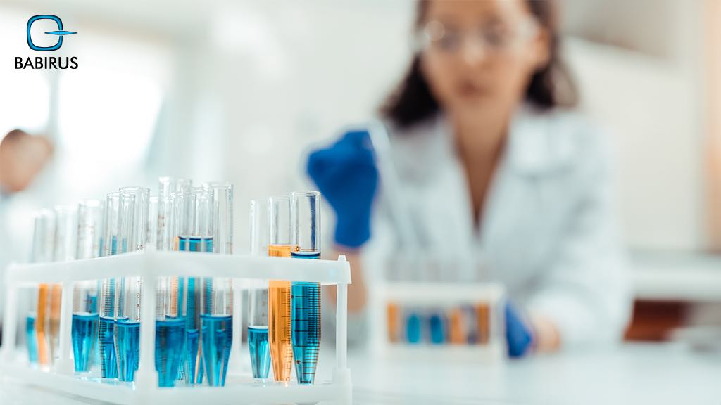
Sample Preparation in Cytopathology

When you think of sample preparation in cytopathology consider it like setting the stage for a performance where each cell has a role to play, thus, this is a crucial first step that brings cellular details clearer under the microscope.
Moreover, when sample preparation is done well, it allows cytologists to see cell structures clearly with all the required details, leading to the accurate diagnosis of infections, cancers, and other abnormalities.
In this article, we will walk through the techniques for collecting and preparing cytopathology samples, and the factors that affect their quality.
The Crucial Role of Sample Preparation:
Sample preparation is at the heart of cytopathology, simply because when a sample is not prepared correctly, then even with the most sophisticated techniques the results will be less reliable.
Thus, each step, from preserving cells to avoiding contamination, plays a vital role in making sure the sample truly represents what is happening at the cellular level, leading to good sample preparation in cytopathology that tells the real story and enables cytologists to make early and accurate diagnoses.
The Best 3 Cytopathology Sample Collection Techniques:
The journey of cytology begins with sample-collecting techniques that differ depending on the type of cells needed to be analyzed:
1. Exfoliative Cytology:
This method gathers cells that are naturally shed from body surfaces, like the cervix, mouth, or lungs, and the most well-known example is the Pap smear, which is used to screen for cervical cancer.
2. Fine-Needle Aspiration (FNA):
The FNA technique indicates using a thin needle to collect cells from lumps or masses in areas like the breast, thyroid, or lymph nodes, which is considered a minimally invasive way and offers a focused sample for analysis.
3. Body Fluid Aspiration:
In this technique, fluids are taken from body cavities, such as pleural, peritoneal, or cerebrospinal fluid, to diagnose the carried cells and reveal information about infections or cancer.
Popular Sample Preparation Methods:
After collecting cells, they need careful preparation to keep them safe and ready for analysis:
Smear Preparation:
This is considered the traditional approach among preparation methods, in which the sample is spread thinly across a slide, however, although this method is widely used in cytology, yet sometimes it could lead to uneven cell distribution, which can impact analysis and the results.
ThinPrep:
This liquid-based technique suspends cells in a fluid medium before they are applied to the slide, which allows for a more even cell distribution, reduces contamination, and leads to clearer slides.
Cytospin:
A centrifuge-based method that concentrates cells from fluid samples onto a slide, which is especially useful when working with small cell quantities in body fluids, offering a detailed view for examination.

Staining Techniques in Cytopathology:
Staining is an essential part of sample preparation in cytopathology, as it highlights cellular structures, helping to differentiate normal cells from abnormal ones.
Papanicolaou Stain:
Commonly known as the Pap stain, this is the go-to stain in cytology, it is capable of enhancing cellular details, like nuclei and cytoplasm, and is especially helpful for examining exfoliative cytology samples.
Special Stains:
Based on the condition being investigated, other staining techniques like the Gram stain or acid-fast stain may be applied to identify specific pathogens or cell types, these stains are more than important when more detailed insights are needed, particularly for infections.
Factors That Impact Sample Quality:
Many factors impact the quality of sample preparation in cytopathology, which makes the real difference between a clear diagnosis and inconclusive results:
· Sample Adequacy:
A good sample means having enough cells to achieve a reliable diagnosis, so, if there are not enough, the sample results may be uncertain or even incorrect.
· Preservation:
Keeping cells complete is essential, thus, using fixatives, like alcohol, is important to help preserve them, and ensure samples stay stable and ready for examination under the microscope.
· Contamination:
Elements like blood, mucus, or foreign particles can obscure cell details, complicating the analysis, that is why careful collection and preparation is vital in keeping samples free from contaminants.
Quality Control in Cytopathology:
High-quality control standards are key to achieving accurate, reliable results, particularly when talking about sample preparation in cytopathology.
Therefore, professional laboratories follow to strict protocols to monitor sample adequacy, assess staining effectiveness, and ensure equipment reliability.
Moreover, with quality control in place, every step of the process is closely managed, reducing the chance of errors and improving diagnostic accuracy.
7 FAQs About Sample Preparation in Cytopathology:
Let us share with you the most asked questions and their answers about sample preparation in cytopathology:
1. How Do You Prepare Samples for Cytological Examination?
Samples can be prepared using various methods, such as smear preparation, thin prep cytology, or cytospin, depending on the sample type and the cells under examination.
2. What Is the Difference Between Smear Preparation and Liquid-Based Cytology?
In smear preparation, the sample is directly spread onto a slide, whereas in liquid-based cytology a liquid medium is used to suspend the cells before placing them on the slide, resulting in a more even distribution.
3. What Stains Are Used in Cytopathology?
The most frequently used stain is the Papanicolaou stain, however, there are other special stains like Gram stain or acid-fast stain that are used for more detailed cellular analysis when necessary.
4. How Can I Ensure the Quality of Samples for Cytology?
To maintain sample quality, it is important to collect a suitable number of cells, use proper preservation methods, and avoid contamination during collection and preparation.
5. What Are the Common Errors in Sample Preparation?
Frequent errors in sample preparation in cytopathology include collecting an insufficient sample, insufficient preservation, and contamination, all of which can impact the accuracy of the outcomes and by default the diagnosis.
6. What Are the Quality Control Measures in Cytopathology Laboratories?
Laboratories follow strict protocols to monitor sample suitability, ensure proper staining techniques, and verify that equipment is functioning correctly to maintain high-quality standards.
7. How Can Sample Preparation Errors Affect Cytological Diagnosis?
Any mistake in sample preparation can lead to false negatives or inconclusive findings, potentially delaying diagnosis and treatment, thus, proper handling of samples is crucial to prevent these issues.
Eventually,
Sample preparation in cytopathology is a critical step that ensures the accuracy of diagnoses in everything from routine screenings to cancer detection.
Therefore, accuracy in all the steps is required from collecting samples to analyses based on the right techniques and quality control measures to help cytologists provide reliable and timely results that improve patient care.
