Challenges and Limitations of Cytopathology

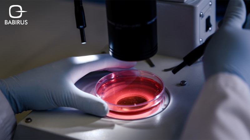
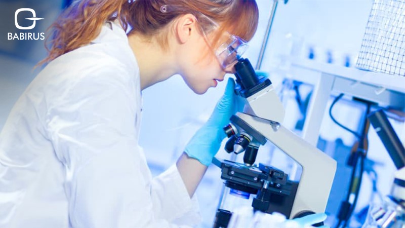
Every cell tells a story, and in cytopathology, the microscope is our window into understanding these narratives, from cancer clues to signs of infections. However, despite its key role in diagnostics, cytopathology challenges persist, impacting diagnostic consistency and accuracy. Factors like sample quality, interpretive differences, and technology limitations can affect cytology’s effectiveness. This blog delves into some primary cytopathology challenges and the limitations cytopathologists face in various applications.
Continue reading as we uncover the core challenges and limitations that shape the practice of cytopathology, revealing the unique obstacles cytopathologists overcome to bring diagnostic precision to a range of applications, as well as answer some frequently asked questions about cytopathology challenges.
Challenges in Sample Acquisition and Preparation
One of the significant cytopathology challenges is related to sample adequacy. For an accurate diagnosis, a cytological sample must contain enough cells and be free from contamination or degradation. However, obtaining high-quality samples can be difficult, especially in cases where cells from specific areas are challenging to reach, such as deep-seated organs or small lesions. For instance, sample limitations in cytology are common when dealing with fine-needle aspirations, where the needle may not always capture sufficient cellular material. Inadequate samples can lead to inconclusive results, requiring further invasive procedures or repeat sampling, which delays diagnosis and treatment.
Interpretational Challenges and Interobserver Variability
Interpretive challenges in cytopathology also contribute to its limitations. Cytological diagnosis often relies on subtle differences in cell appearance, which can vary based on a pathologist’s expertise. Variability among observers, known as interobserver variability, can result in discrepancies in diagnoses. For example, when it comes to cytopathology and cancer, differentiating between benign and malignant cells in early-stage cancer may yield different results among pathologists. Cytopathology also involves subjective interpretation, especially when diagnosing complex conditions, which can impact the consistency of results. Efforts to improve interobserver consistency, such as standardized criteria and additional training, aim to reduce these cytopathology challenges.
Limitations of Traditional Cytology Techniques
While technology has advanced significantly in recent years, certain technological limitations in cytopathology remain. Traditional microscopy, while foundational, has limitations in terms of resolution and contrast, which may hinder the ability to detect subtle cellular changes. Additionally, the manual nature of traditional cytological analysis can be time-consuming and prone to human error. Digital imaging like the Lituo LTS-BL1000 Digital Slide Scanning & Imaging System, help overcome these limitations by enabling rapid slide scanning, automatic focusing, and precise imaging. Equipped with an AI-powered identification system and a robust database, the Lituo slide reader enhances diagnostic efficiency and accuracy, allowing cytopathologists to manage large volumes of samples effectively. Such advancements in digital imaging provide a pathway to overcoming traditional limitations and improving the reliability of cytological diagnoses.
Cytology Limitations in Specific Applications
When it comes to specific applications like cancer and infectious diseases, cytopathology challenges present unique obstacles. Cytopathology and cancer is a field where early-stage diagnosis can be particularly difficult due to the subtle changes in cells at the initial stages. Detecting precancerous or very early malignant changes requires highly sensitive tools and interpretation, which may not always be feasible in routine cytology labs.
When it comes to cytopathology and infectious diseases, certain pathogens may not present clear cytological markers or may resemble benign cellular changes, complicating the identification process. Infectious agents like viruses or fungi may be difficult to distinguish using traditional staining techniques alone, and in some cases, molecular methods are necessary to confirm the diagnosis. These specific application challenges underline the importance of integrating cytology with other diagnostic methods, such as molecular testing, to improve accuracy.
FAQs
To deepen our understanding of cytopathology challenges, let’s look at some frequently asked questions that explore key issues and effective approaches.
What are the common challenges faced in obtaining adequate samples for cytological examination?
Obtaining adequate samples can be challenging when targeting hard-to-reach areas or small lesions, leading to samples with insufficient cellular material or contamination, which affects diagnostic accuracy.
How can we improve the interobserver variability in cytopathology?
Standardized diagnostic criteria, additional training, and employing digital imaging tools are some approaches to improve consistency among cytologists in interpreting cytological samples.
What are the limitations of traditional microscopy techniques in cytology?
Traditional microscopy has limitations in resolution and contrast, which can make detecting subtle cellular abnormalities more difficult. Digital imaging and AI are being explored to address these limitations.
How can artificial intelligence be used to address challenges in cytological interpretation?
AI can assist in identifying patterns in cytological samples and may help automate parts of the diagnostic process, potentially increasing consistency and accuracy in cytology.
What are the challenges of using cytopathology for early-stage cancer detection?
Early-stage cancer detection requires identifying subtle cellular changes that may be difficult to detect with standard cytology. Enhancements in molecular and digital tools could improve early detection.
How can we improve the accuracy of cytology in diagnosing infectious diseases?
Incorporating molecular methods and specialized staining techniques can enhance accuracy in identifying infectious pathogens in cytology samples, providing a clearer view of the cellular structures affected.
What are the future trends in cytopathology to overcome current limitations?
Future trends in cytopathology like digital cytology, AI integration, and advanced molecular techniques are likely to improve diagnostic accuracy, efficiency, and early detection capabilities in cytopathology.
What are the ethical considerations in addressing challenges in cytopathology?
Ensuring patient consent for advanced testing methods, maintaining accuracy in AI-assisted diagnostics, and addressing accessibility issues are key ethical considerations.
To conclude,
While cytopathology challenges still exist, ongoing advancements in sampling methods, technology, and diagnostic tools are gradually overcoming key limitations. From improving sample adequacy to boosting diagnostic accuracy, these developments are strengthening cytopathology’s role in identifying diseases such as cancer and infections. With each innovation, cytopathology becomes better equipped to support precise and timely diagnoses that improve patient care.

